Residues in contact with ligands in PDB files
2
1
Entering edit mode
4.1 years ago
lumal29 ▴ 80

Hi everyone,

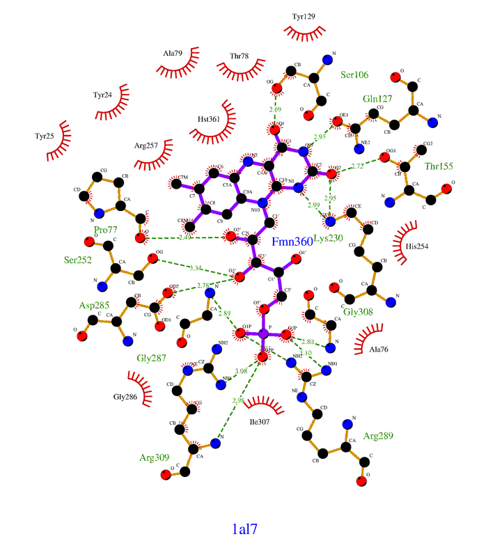
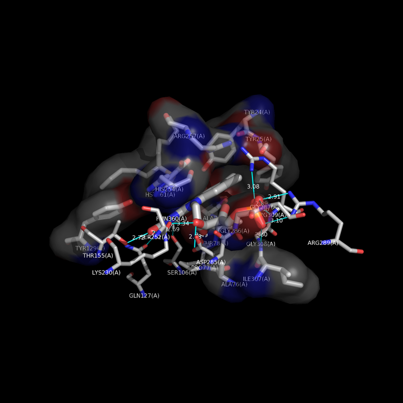
I am trying to found out what residues are in contact with a ligand (the binding site) in a PDB file. To give you an example, I need to find the residues in contact with FMN ligand in the PDB entry 1AL7.

First of all, can we find this type of information in a PDB file? Or do we need a software to calculate it? I think the first question is yes, but I am not sure: I used the new mmCIF format, as they are well organized compared to the old version. In this format, there is a Data Category called "chem_comp" and the dictionnary on the PDB website says :

"Data items in the CHEM_COMP category give details about each of the chemical components from which the relevant chemical structures can be constructed, such as name, mass or charge." It allows me to know if there is the presence of a FMN ligand.

Another category is "struct_site":

"Data items in the STRUCT_SITE category record details about portions of the structure that contribute to structurally relevant sites (e.g. active sites, substrate-binding subsites, metal-coordination sites)."

In my example, there is a line "AC1 Software ? ? ? ? 22 'BINDING SITE FOR RESIDUE FMN A 360'"

And then, you have another category called "struct_site_gen":

"Data items in the STRUCT_SITE_GEN category record details about the generation of portions of the structure that contribute to structurally relevant sites."

This category is, I guess, my answer, because I see all the residues with their number sharing the same id (AC1) than in struct_site.

Can I use this information for the binding site or you guys think I have to use a software to calculate the residues in the binding site? If so, which one?

Thank you

PDB protein residues ligand binding site • 2.2k views
ADD COMMENT
0
Entering edit mode

Hello, I was wondering if you had success in finding the non-bonded contact residues programatically? I am performing similar research and would greatly appreciate if you could share any success you have had regarding this task!

ADD REPLY
1
Entering edit mode
4.1 years ago
jgreener ▴ 390

Yes, you can use the information in STRUCT_SITE_GEN listed under AC1 to get the binding residues: TYR24, TYR25, ALA76 etc.

If you did want to do this from the coordinate data, one way would be to use BioStructures.jl in Julia:

using BioStructures
struc = read("1AL7.cif", MMCIF)
res = collectresidues(struc, standardselector)
for r in res
    if distance(r, struc["A"]["H_360"]) < 4.0
        println(r)
    end
end

This returns more or less the same residues as STRUCT_SITE_GEN. You could also do this with Biopython.

ADD COMMENT
1
Entering edit mode

Thank you very much! I don't know Julia very much, just learned the basics, but I will take a look at it, BioJulia looks pretty interesting! I have few questions though:

  • I guess in the if statement, you select the chain A and the residue 360 which in fact is FMN ligand, but how do you know it's 360? If I have several pdb files, how can I find it programmaticaly?
  • Is 4 for 4 angstrom? If so, Why 4? Thanks again!
ADD REPLY
0
Entering edit mode

You could find it programmatically by finding the first residue with the desired residue name:

allres = collectresidues(struc)
fmnres = allres[findfirst(r -> resname(r) == "FMN", allres)]

Yes, 4 is for 4 Angstrom. I just chose it as a distance below which atoms could be considered as contacting, but you could use any value.

ADD REPLY
1
Entering edit mode
4.1 years ago
Mensur Dlakic ★ 27k

I highly recommend LigPlot+. It is easy to use and intuitive. It will produce schematic diagrams in PostScript, and has a direct interface that will connect with PyMol if you have it installed. For your structure, it will make the two images below.

enter image description here

enter image description here

ADD COMMENT
0
Entering edit mode

Thank you very much! I heard about Ligplot before, but it's not what I was looking for, as I have to find the residues programaticaly for a several hundreds of pdb files. I would have a question though: If I understand correctly, the residues with a green dash line are supposed to be in contact with FMN, but what about the residues in red (eyelash)?

ADD REPLY
0
Entering edit mode

The explanation is below the image in the LigPlot+ web page: Hydrogen bonds are shown as green dotted lines, while the spoked arcs represent protein residues making nonbonded contacts with the ligand.

ADD REPLY

Login before adding your answer.

Traffic: 2608 users visited in the last hour
Help About
FAQ
Access RSS
API
Stats

Use of this site constitutes acceptance of our User Agreement and Privacy Policy.

Powered by the version 2.3.6